
IMPLANT AND SURGERY
Bone Powder (Graft)
A procedure that restores bone that is insufficient for an implant
Provided at our clinic in Altunizade, Üsküdar (Istanbul).
IN SHORT
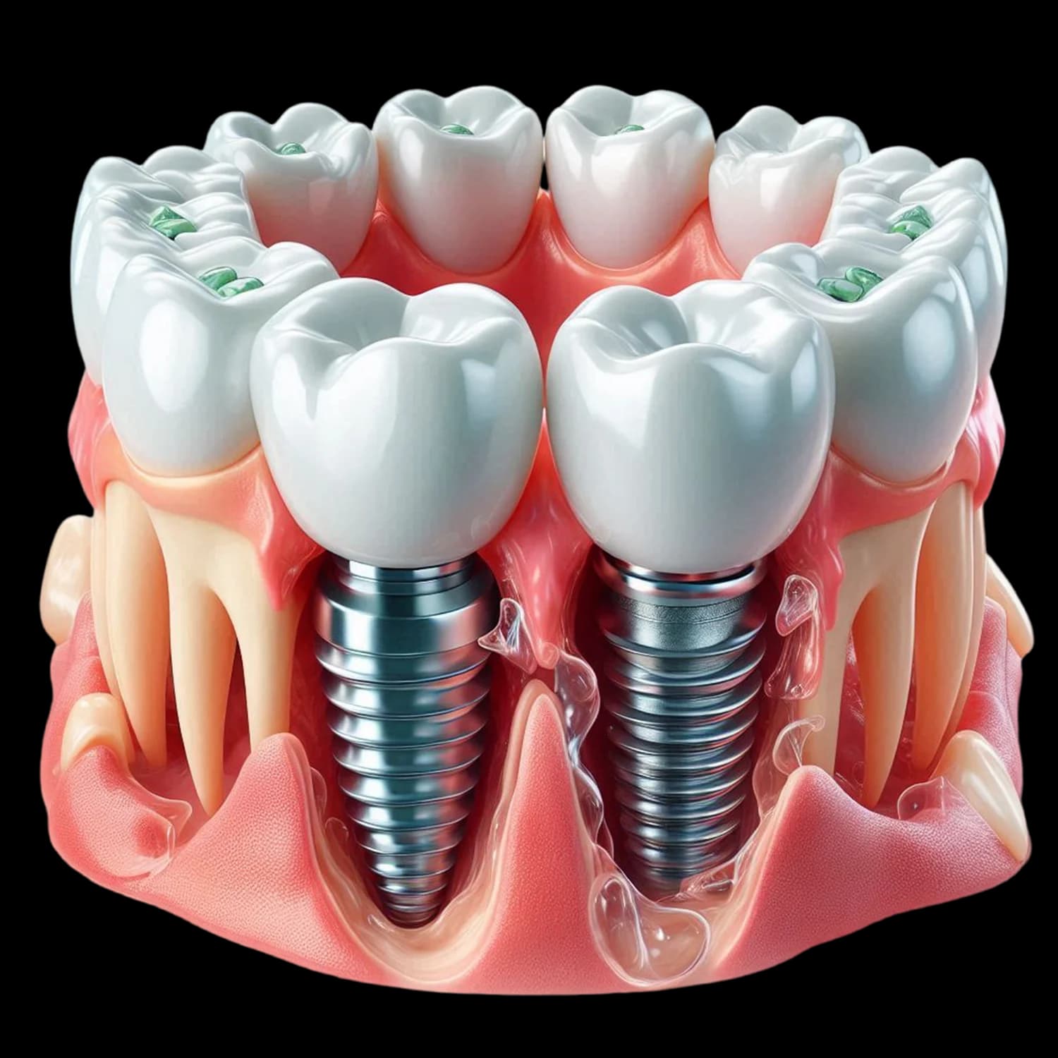
A bone graft is a procedure applied when there is not enough volume in the jawbone for an implant. Graft material is placed in the deficient area and forms a scaffold for the body's own bone to grow into it. The graft material itself does not turn into bone; the new bone is the patient's own tissue. Healing usually takes 4–9 months. At the end of this period, a bone volume into which an implant can be placed is formed.
Why does bone resorb?
During chewing, the tooth root transmits force to the surrounding bone. This force is the stimulus that keeps the bone in existence.
When the tooth is extracted, this stimulus disappears and the bone in that area begins to resorb. Resorption is seen fastest within the first 6–12 months after extraction. The loss in the width of the bone is greater than the loss in its height.
The practical consequence of this is: when an implant is considered years after a tooth has been extracted, the bone has usually become insufficient. The longer the time between the extraction and the implant, the greater the need for a graft.
Other causes of bone resorption: advanced gum disease, long-term use of a removable denture, trauma and pathologies such as cysts.
How does a graft work?
This is the subject patients most often misunderstand.
The graft material does not turn into bone. The material placed in the gap forms a scaffold (a base) for the body's own bone cells to grow into it and make new bone. Over time the graft material is partly or fully resorbed and is replaced by the patient's own bone.
In other words, the graft is not bone; it is the place where bone will form. This process takes time. Healing usually takes 4–9 months, depending on the size and type of the graft.
What is a membrane?
The graft material is usually covered with a thin sheet called a membrane.
Its purpose is this: the soft tissue (the gum) heals much faster than the bone. Without a membrane, the soft tissue fills the gap where the graft is and leaves no room for bone to form. The membrane keeps the soft tissue out and buys time for the bone.
Some membranes resorb on their own; others need to be removed.
When is it applied?
At the same time as extraction (socket preservation). The graft is placed in the gap while the tooth is being extracted. Because it is done before the bone resorbs, it is the most effective approach. This option should be considered when extracting a tooth for which an implant is planned.
At the same time as the implant. If the bone deficiency is limited, a graft can be placed in the gap around the implant while it is being placed. No additional surgical session is needed.
As a separate stage before the implant. If the bone deficiency is pronounced, the graft is done first, healing is awaited (4–9 months), and the implant is then placed. The treatment time is longer.
Who is it not suitable for? In which cases does the risk increase?
Heavy smoking. It is one of the most common causes of graft failure. Smoking directly impairs the blood circulation and healing in the graft area.
Uncontrolled diabetes.
Use of bisphosphonate-type medication or a history of radiotherapy to the jaw region. Assessment together with your doctor and the relevant specialist is required.
Active infection in the area. It must be resolved first.
Untreated gum disease.
Patients whose oral care is not sufficient.
The procedure and healing
Examination and assessment with a panoramic X-ray
Where the extent of the bone deficiency and its relationship with the neighbouring anatomical structures need to be clarified, three-dimensional imaging is requested.
Planning
The type and amount of graft, and whether it will be done at the same time as the implant, are determined.
Surgery
The procedure is carried out under local anaesthesia. In patients with a high level of anxiety, the treatment sessions can be divided and breaks can be taken.
Healing
Usually 4–9 months. During this period no force should be placed on the area.
Assessment and implant
Bone formation is assessed radiographically; if it is sufficient, the implant is placed.
Risks and possible complications
Failure of the graft to take. New bone may not form sufficiently; the procedure may need to be repeated.
Infection.
Exposure of the membrane. If the gum opens and the membrane becomes visible, the risk of graft loss increases.
Swelling, bruising and pain. These are expected findings.
Loss of part of the graft material.
Smoking markedly increases all of these risks. Stopping or minimising it throughout the healing period is recommended. In the first days, swelling, bruising and tenderness are expected findings; no pressure should be placed on the area.
Types of graft
Materials from different sources are used for a graft. Which type is used is determined according to the size of the deficiency, the area and the patient's condition. Often more than one type is used together. The table below compares the four types.
Compare the options
| Source | Property | Additional procedure | |
|---|---|---|---|
| Autograft | The patient's own bone (usually taken from the jaw) | As it is the patient's own tissue, no compatibility problem is expected. | A second surgical site is required. |
| Allograft | Processed tissue of human origin | No additional surgical site is required. | — |
| Xenograft | Processed tissue of animal origin (usually bovine) | Resorbs slowly; preserves the volume for a long time. | — |
| Alloplast | Synthetic material | Contains no biological source. | — |
Which option is suitable is decided together after an intraoral examination.
Frequently Asked Questions
The graft material itself does not turn into bone. The material forms a scaffold for the body's own bone cells to grow into it and make new bone. Over time the graft material is partly or fully resorbed and is replaced by the patient's own bone. In other words, the graft is not bone; it is the place where bone will form.
It usually takes 4–9 months, depending on the size and type of the graft. At the end of this period, bone formation is assessed radiographically; if it is sufficient, the implant is placed.
It varies according to the type of graft used. There are four types: the patient's own bone (autograft), processed tissue of human origin (allograft), processed tissue of animal origin (xenograft — usually bovine) and synthetic material (alloplast). If you have a preference on this matter, you need to state it before treatment; suitable alternatives are assessed.
Insufficient bone does not mean an implant definitely cannot be done. Bone volume can be increased with a graft. This lengthens the treatment time. How insufficient the bone is, and whether a graft can be applied, are determined after an examination and radiographic assessment.
This option should be considered when extracting a tooth for which an implant is planned. A graft at the same time as extraction (socket preservation) is the most effective approach, because it intervenes before the bone begins to resorb. The longer the time between the extraction and the implant, the greater the need for a graft.
Smoking directly impairs the blood circulation and healing in the graft area; it is one of the most common causes of graft failure. It does not mean treatment cannot be done, but the success rate falls markedly. Stopping or minimising it throughout the healing period is recommended.
The procedure is carried out under local anaesthesia and no pain is felt during it. Afterwards, swelling, bruising and tenderness lasting a few days are expected findings; they can be kept under control with the medication your dentist recommends.
Related treatments
The content on this site is for information only and does not replace a clinical examination. The suitability, duration and scope of treatment are determined after a clinical assessment.
This content was prepared by Dt. Mevlüt Yazıcı.
Request an appointment.
Fill in the form to request an appointment and we'll get back to you shortly. You can also call us directly or message us on WhatsApp.
Altes Diş Kliniği
Altunizade, Üsküdar, İstanbul
- PHONE+90 533 474 43 94
- EMAIL[email protected]
- ADDRESSAlperen İş Merkezi, Valide-i Atik Mahallesi, Altunizade, Nuhkuyusu Cd D:191/B, 34664 Üsküdar/İstanbul
- WORKING HOURSWeekdays 10:00–19:00 · Saturday 10:00–16:00 · Sunday closed